

Eye Care Resources
Optic Neuritis in North York: When Vision Looks Dim or Colours Fade
April 11, 2025
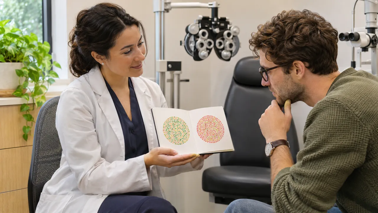
Sometimes a vision problem is difficult to describe as blur. One eye may seem to see through dimmer light. A red jacket may look less vivid, or a familiar room may appear slightly washed out even though the lighting has not changed. These observations matter, especially when they are new or accompanied by discomfort when the eye moves.
Optic neuritis is one possible explanation for a change in brightness, colour or vision. It is not something to diagnose by comparing colours on a phone screen. People in North York experiencing a new reduction in sight should seek prompt professional assessment so that the cause and urgency can be established.
If optic neuritis has already been mentioned at an appointment, there may be several unfamiliar next steps: a specialist referral, an MRI, blood tests or a discussion of treatment. Knowing what each step is meant to answer can make the process less overwhelming without trying to predict the result in advance.
What the optic nerve does
The optic nerve carries visual signals from the eye to the brain. Optic neuritis means inflammation affecting that nerve. It can interfere with clarity, brightness, colour and areas of the visual field. Pain with eye movement is common, although the exact symptoms vary. The North American Neuro-Ophthalmology Society explains optic neuritis and its effects.
A useful distinction is between focusing light and transmitting visual information. A glasses prescription helps the eye focus, but a nerve problem requires a different kind of investigation. This is why an examiner may continue testing even when changing lenses does not restore the expected clarity.
Avoid treating the word neuritis as a complete explanation of the cause. It describes inflammation. The clinical team still needs to consider why it has occurred and whether the pattern fits the expected diagnosis. A discussion of possibilities is part of that investigation, not a declaration that every possibility applies to you.
Describe the change in ordinary language
You do not need medical vocabulary to give a useful history. Explain when you first noticed the difference, whether it affects one or both eyes, and how it has changed since then. If the vision seems darker, say darker. If colours are less vivid, describe an example that is easy to remember.
Mention discomfort with looking up, down or sideways, but do not repeatedly move the eyes to provoke pain. Tell the clinician about other symptoms, recent illness, previous episodes and known neurological or autoimmune conditions. Bring a medication list, including recent changes.
Try to distinguish the beginning of the problem from the day you became certain it was present. For example, you might have noticed difficulty reading on Monday and a brightness difference on Wednesday. That timeline is more helpful than choosing one date and leaving out the earlier change.
If you made notes before the visit, keep them short. A few clear observations are easier to discuss than a large collection of home test results. Screens, room lighting and camera settings can all vary, so repeated digital comparisons should not replace an examination.
New loss of vision needs prompt assessment
A new reduction in vision, especially with eye pain or a marked difference between the eyes, should be assessed urgently. Contact an eye care professional and describe the symptoms directly. If timely assessment is unavailable, use an appropriate urgent medical service. Do not wait for a routine glasses appointment simply because the eye looks normal from the outside.
Sudden severe vision loss or a visual change accompanied by weakness, speech difficulty, severe headache or other neurological symptoms warrants emergency care. There are causes of acute visual loss other than optic neuritis, and the priority is timely evaluation rather than deciding which diagnosis seems most familiar.
Avoid driving when the change affects your ability to see safely. Ask someone to accompany you if practical, particularly when dilation or further hospital assessment may be needed. Gathering every old record is less important than getting a new significant symptom assessed promptly.
Our emergency eye care information explains how to contact the clinic about urgent concerns. A routine online appointment request is not an emergency response system, so call or seek urgent care directly when symptoms are sudden or worsening.
Why the examination includes more than an eye chart
The assessment may compare visual acuity, colour vision, pupil responses and peripheral vision, as well as the appearance of the optic nerve. The optic nerve can be inflamed behind the eye even when its visible surface is not obviously swollen. MedlinePlus describes the examination and investigations used for optic neuritis.
Ask what each result contributes. Reading letters measures one aspect of vision, while colour and field testing can reveal difficulties that a letter score alone does not capture. If you can read a small line but still find everyday vision noticeably different, explain that rather than assuming the experience must be unimportant.
Some tests require concentration. Tell the technician if you are unsure about the instructions or need a brief pause. A visual field test is not an exam you pass by guessing correctly. Following the instructions comfortably helps produce a result the clinician can interpret.
Keep copies of reports when your care involves different offices. Our guide to visual field testing explains that appointment in more detail. Ask whether the specialist will receive the images and results automatically or whether you need to authorize a transfer.
What a specialist referral is intended to clarify
A neuro-ophthalmologist considers the connection between the visual system and the nervous system. Referral may help distinguish optic neuritis from other causes of optic nerve dysfunction. MRI or additional tests may be used to investigate the nerve and possible associated conditions. Moorfields describes the role of neuro-ophthalmology.
Receiving a referral does not tell you the eventual diagnosis. Ask what question the referring clinician wants answered and whether any investigation should happen before the appointment. Make sure the receiving office has the correct telephone number and knows about a significant change in symptoms.
If an MRI is ordered, ask the imaging team about preparation and any information they need regarding implants, pregnancy, allergies or other medical considerations. Do not decide that a scan is unnecessary because the eye feels better by the time the appointment arrives. Ask the ordering clinician before changing the plan.
Confirm how results will be communicated. A completed test and a reviewed test are separate steps. Knowing who will contact you, and what to do if you have not heard by the expected time, helps prevent uncertainty from becoming an avoidable gap in care.
A discussion of multiple sclerosis is not a diagnosis
Optic neuritis can occur with multiple sclerosis, other inflammatory disorders, infections or without an identified associated condition. Not everyone who experiences optic neuritis has or will develop multiple sclerosis. Individual assessment and imaging help clinicians interpret the situation; a general risk figure cannot answer the question for one person.
It is reasonable to feel concerned when a possible neurological condition enters the conversation. Ask which findings support the current concern, which remain uncertain and what investigation can clarify them. Write down the distinction between a confirmed diagnosis and a possibility being checked.
Avoid comparing your symptoms with an online account and assuming that the entire course will be the same. The term optic neuritis includes different clinical circumstances. Someone else’s recovery time or treatment may not be a useful guide to your own needs.
If uncertainty is affecting sleep or daily functioning, tell your care team. You can ask for a clearer explanation, accessible written information or support while investigations proceed. Understanding what is known today is often more manageable than trying to solve every possible future outcome at once.
Treatment decisions depend on the cause and pattern
Some typical episodes improve without treatment, while corticosteroids may be used to speed recovery in selected situations. Other causes of optic nerve inflammation need different or more intensive treatment. The decision belongs with the treating specialist after considering the examination, investigations and overall medical history.
Do not use leftover steroid tablets or eye drops to treat suspected optic neuritis. The appropriate treatment, route and dose cannot be inferred from the fact that a medicine is described as anti-inflammatory. Ask what the proposed treatment is intended to achieve and which side effects require contact.
If treatment is prescribed, obtain written instructions and identify the team responsible for questions. Ask whether your family physician needs to monitor any aspect of general health. Make sure the clinician knows about other medicines and relevant conditions before treatment begins.
A useful follow-up discussion includes both the next examination and the signs that should bring you back sooner. Improved comfort does not necessarily answer every question about visual recovery. Equally, a slow change should be discussed in the context of your results rather than judged against a fixed online timetable.
Plan around the vision you currently have
While assessment or recovery is underway, adjust tasks according to what feels reliable. Increase text size, use comfortable lighting and allow more time for reading important information. If colour discrimination is affected, avoid relying only on colour to identify medication, work materials or warning signals.
Talk with your clinician about driving and job duties that require precise visual judgement. For commuting in North York, arranging a ride or accompaniment may be sensible when depth, contrast or clarity is uncertain. Do not use confidence on a familiar route as proof that driving is safe.
Tell family members what support is helpful. You might welcome help getting to a specialist appointment while preferring to manage your own notes and calls. Clear requests preserve independence and reduce the burden of explaining the situation repeatedly.
After urgent assessment, keep the recommended specialist follow-up and book routine eye care according to the plan. Report a new or worsening change promptly, including after an earlier episode has improved. The most useful approach is a clear symptom history, completed investigations and a care team that knows what has changed.