

Eye Care Resources
Ocular Hypertension in North York: What High Eye Pressure Means
September 1, 2026
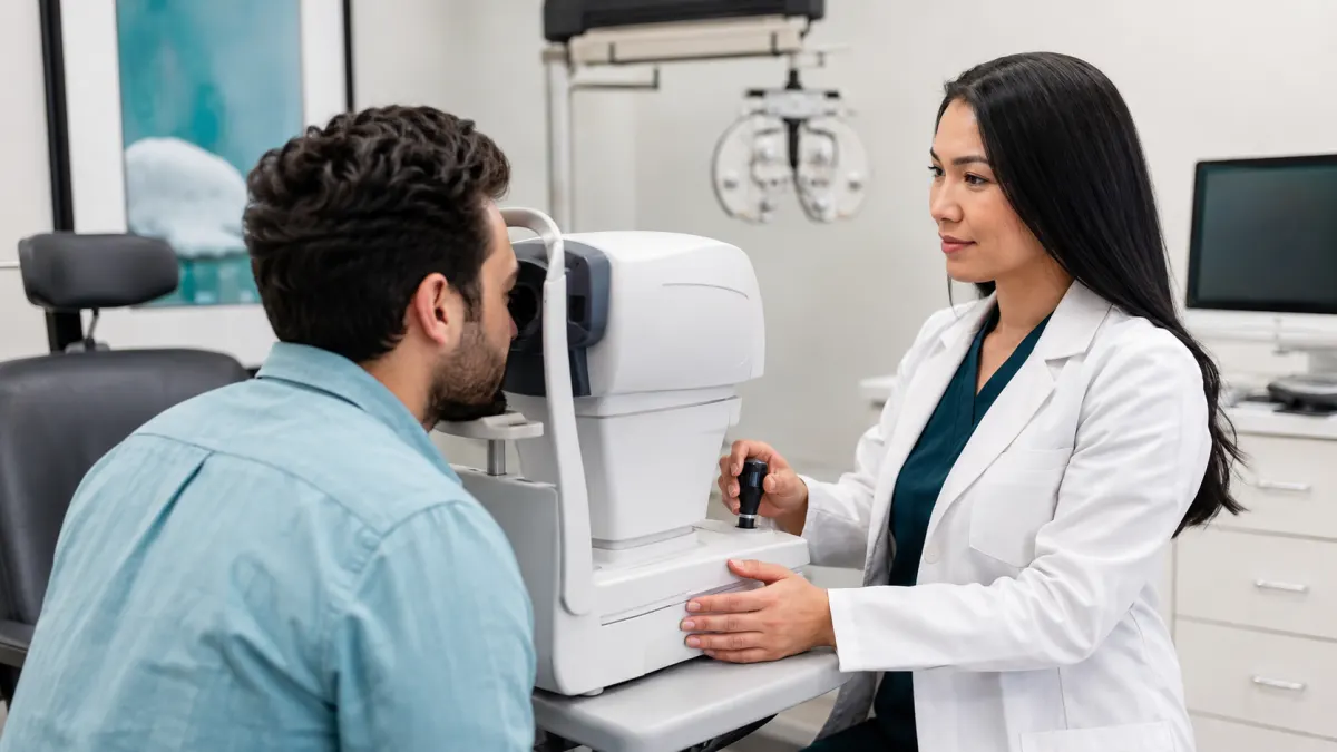
An eye-pressure reading comes back higher than expected, yet vision is clear and the optic nerves look healthy. Does that mean glaucoma? Not necessarily. It may be ocular hypertension: pressure inside the eye is elevated, but the characteristic optic-nerve damage and visual-field loss of glaucoma have not been found.
Ocular hypertension is not simply “glaucoma that has not been noticed yet,” nor is it a result that can be ignored. Higher pressure increases glaucoma risk, but people tolerate pressure differently. Some develop optic-nerve damage at readings considered statistically normal, while others maintain healthy nerves despite higher measurements.
The purpose of assessment is not to chase one universal number. It is to estimate an individual’s risk, establish reliable baselines, look for change, and decide whether observation or pressure-lowering treatment offers the better balance.
Eye pressure comes from fluid balance
Clear fluid called aqueous humour is continually produced inside the front of the eye. It circulates through the pupil and normally drains through tissue at the angle where the iris and cornea meet. Pressure reflects the balance between production and outflow.
This fluid is different from tears, which coat the outside surface. Watery eyes do not “release” internal eye pressure, and drinking less water is not a treatment for ocular hypertension.
When outflow becomes less efficient, pressure may rise. Anatomy, age, genetics, medications, inflammation, trauma, pigment, and other ocular factors can influence the drainage system. Most people with ocular hypertension feel nothing; the condition is usually discovered during an examination.
Ocular hypertension and glaucoma are different findings
Glaucoma is a group of diseases that damage the optic nerve and can cause permanent visual-field loss. Eye pressure is an important modifiable risk factor, but it is not the definition of glaucoma.
Ocular hypertension generally means pressure is higher than expected without detectable glaucomatous nerve damage. A glaucoma suspect may have elevated pressure, a suspicious optic-nerve appearance, a strong family history, an unusual visual field, or another risk feature without enough evidence for a definite diagnosis.
These labels can change as more information is collected. A single normal field does not prove lifelong safety, and one unreliable field does not establish glaucoma. Diagnosis depends on the pattern and change across time.
The “normal range” is not a personal safety line
Eye pressure is measured in millimetres of mercury. Many references describe a population range around the low teens to low twenties, but a cutoff such as 21 mmHg is not a pass-fail boundary. It is possible to have pressure above that level without damage or to have glaucoma below it.
Clinicians interpret the reading with corneal thickness, measurement method, time of day, optic-nerve structure, fields, age, family history, ancestry, prescription, and other health factors. The target, if treatment is started, is individualized and may change when new evidence appears.
Avoid comparing your number with a friend’s. Two people at the same measured pressure can have different corneas, nerves, histories, and risk.
How pressure is measured
Non-contact tonometry uses a brief air pulse and does not touch the eye. It is useful for screening and can provide reliable information in appropriate circumstances. Applanation tonometry estimates pressure by measuring the force required to flatten a small area of the anesthetized cornea. Handheld and rebound devices use other methods.
Readings vary somewhat among instruments and even between repeated measurements. Holding the breath, squeezing the eyelids, leaning awkwardly, a tight collar, and poor positioning can affect results. Try to breathe normally, keep the face relaxed, and let the clinician support the lids if needed.
An unexpected result is often rechecked. That does not mean the first number was meaningless; it means pressure is a biological measurement with context, not a fixed label.
Corneal thickness changes interpretation
Tonometry measures through the cornea, so corneal properties influence the estimate. A thicker central cornea may contribute to a higher measured value, while a thinner cornea may lead to underestimation and can also be associated with greater glaucoma risk in some contexts.
Pachymetry measures central corneal thickness. The result helps interpret pressure and risk, but there is no simple calculator that perfectly converts every reading into a “true pressure.” Corneal biomechanics and instrument type also matter.
Previous laser refractive surgery can alter corneal thickness and curvature, complicating comparison with old readings. Tell the clinic about LASIK, PRK, other corneal surgery, or keratoconus, and bring historical records when available.
Gonioscopy examines the drainage angle
The angle where fluid leaves the eye cannot be fully assessed by looking straight through the cornea alone. Gonioscopy uses a mirrored diagnostic lens on the anesthetized eye to view this anatomy. It helps determine whether the angle is open, narrow, scarred, or affected by pigment, inflammation, trauma, or new blood vessels.
Open-angle and angle-closure mechanisms are managed differently. A narrow angle may carry a risk of sudden closure in particular eyes, while chronic open-angle disease usually progresses silently.
Ask whether the drainage angle has been assessed if pressure is elevated or dilation safety is being considered. New severe eye pain, redness, halos, headache, nausea, or vomiting is not routine ocular hypertension; it can signal acute angle closure and requires emergency care.
The optic nerve must be examined and documented
The optic nerve contains the fibres carrying visual information to the brain. Glaucoma damages these fibres in characteristic patterns. The examiner evaluates the nerve rim, cup, size, asymmetry, colour, nearby retinal nerve-fibre layer, and possible hemorrhage.
A large cup is not automatically glaucoma. Large healthy nerves often have large cups, while a small nerve can sustain damage without looking dramatically cupped. Comparison with photographs or scans over time is often more informative than an isolated description.
Optical coherence tomography measures retinal nerve-fibre and ganglion-cell layers. It can reveal structural change, but the result is compared with a reference database and must be checked for scan quality, anatomy, prescription effects, and segmentation errors.
Visual fields test function
Automated perimetry asks the patient to respond to dim lights in different locations while looking at a central target. It maps sensitivity rather than how sharp the central chart appears. Early glaucoma can affect peripheral or paracentral areas while ordinary acuity remains excellent.
The test is challenging because it measures threshold perception and requires concentration. False responses, missed fixation, fatigue, dry eye, and an incorrect trial lens can reduce reliability. A suspicious first result is often repeated to see whether the pattern is reproducible.
There is no advantage to guessing. Blink normally, pause when allowed, and click only when a light is seen. Tell the technician if the eye patch, lens, or posture is uncomfortable.
Risk is built from several factors
Features that may influence concern include:
- higher or repeatedly elevated pressure;
- thinner central cornea;
- older age;
- a close family history of glaucoma;
- suspicious optic-nerve structure;
- reproducible visual-field findings;
- certain ancestry-related population risks;
- strong nearsightedness or other ocular anatomy;
- previous eye trauma or inflammation;
- long-term or potent steroid exposure; and
- evidence of change on photographs, OCT, or fields.
No list can determine an individual’s outcome. Some factors are not modifiable, while pressure can often be lowered. The clinician combines findings rather than treating each checkbox independently.
Steroids can raise pressure
Steroid eye drops, injections, tablets, inhalers, nasal sprays, skin creams near the eyes, and other routes can raise intraocular pressure in susceptible people. The response may appear after treatment begins and may be silent.
Do not stop a medically necessary steroid abruptly. Tell the prescribing clinician and eye-care provider about every steroid product, including creams and inhalers. Monitoring can be arranged and treatment adjusted when appropriate.
Never use leftover steroid eye drops for redness. Besides pressure effects, steroids can worsen certain infections and delay healing.
Observation may be the right plan
Not every person with ocular hypertension needs immediate drops. When the estimated short- and long-term risk is low, careful observation can avoid cost, side effects, inconvenience, and treatment burden. Observation is active care, not neglect.
A monitoring plan specifies when pressure, optic nerves, OCT, photographs, gonioscopy, pachymetry, and visual fields will be reviewed. Intervals depend on risk and stability. Keeping appointments matters because glaucoma-related damage is irreversible and often symptomless early.
Try to have follow-up performed with comparable methods and good-quality baselines. Moving clinics does not have to interrupt continuity; request copies of prior fields, OCT reports, nerve photographs, pressures, and corneal thickness.
Treatment is chosen when benefit outweighs burden
Pressure-lowering eye drops reduce fluid production, improve outflow, or both. Different classes have different dosing, side effects, systemic considerations, and storage needs. Laser trabeculoplasty may be an option for selected open-angle cases. Surgery is generally reserved for situations requiring greater pressure control or when other methods are insufficient.
The decision to treat may reflect higher predicted risk, very elevated pressure, suspicious testing, limited ability to monitor, expected lifetime risk, or documented change. A target pressure is an estimate designed to reduce risk, not a guarantee.
If drops are prescribed, demonstrate how you use them. One drop is enough. Close the eye gently rather than squeezing, and ask about pressing at the inner corner to reduce drainage into the nose. Separate multiple drops by the interval instructed so one does not wash out the other.
Side effects and adherence should be discussed honestly
Redness, stinging, dryness, eyelid-skin change, lash growth, iris darkening, fatigue, breathing effects, or heart-rate effects can occur depending on the medication. Report problems rather than silently stopping or rationing drops.
Adherence is difficult when a condition has no symptoms. Link dosing to a stable daily routine, use reminders, and plan refills before travel. Tell the clinic about cost, dexterity, tremor, arthritis, memory, caregiving, or schedule barriers. A plan only works if it is realistic.
Bring all bottles to follow-up. Similar cap colours or generic names can create confusion, and the actual regimen is more useful than a remembered description.
Lifestyle supports health but does not replace monitoring
Regular activity, blood-pressure care, diabetes management, not smoking, and balanced overall health are valuable. They do not substitute for pressure assessment or reverse established optic-nerve damage.
Avoid unproven supplements marketed to “detox” eye pressure. Some products interact with medications or create false reassurance. Large rapid fluid intake, breath-holding strain, prolonged inverted positions, and certain activities can transiently influence pressure, but everyday advice should be individualized rather than based on fear.
Continue exercise unless your clinician identifies a specific restriction. Ask before changing a prescribed medication because of something read online.
Symptoms do not reliably track chronic pressure
Chronic open-angle pressure elevation usually does not cause an ache that rises and falls with the tonometer number. Headache, dry eye, sinus symptoms, and migraine are common and can coexist. Feeling normal also does not prove the pressure or optic nerve is stable.
Acute angle closure is the exception: severe pain, redness, blur or halos, headache, nausea, and vomiting can occur with a sudden pressure rise. Seek emergency care immediately for that pattern.
For most ocular-hypertension patients, scheduled objective testing—not daily sensation—guides management.
Ocular-hypertension care in North York
One pressure number begins a question; it does not finish the diagnosis. Reliable repeat measurements, corneal thickness, angle anatomy, optic-nerve assessment, OCT, photographs, visual fields, history, and change over time create the fuller picture.
Optima Eye Care provides comprehensive examinations and glaucoma-oriented monitoring for North York patients. Bring family history, medication list, prior surgery information, and previous scans or fields when available. To review elevated pressure or establish a baseline, request an eye examination. For sudden severe eye pain, redness, halos, nausea, or vision loss, seek emergency care immediately.