

Eye Care Resources
Keratoconus in North York: Early Signs, Corneal Mapping, and Treatment Options
June 11, 2026
A glasses prescription that changes again and again can be frustrating. Words may still have a faint shadow after the lenses are updated. Headlights stretch into streaks, subtitles appear doubled, and one eye never seems as crisp as the other. Sometimes these changes are ordinary refractive shifts. Sometimes they are clues that the front surface of the eye is becoming irregular.
Keratoconus is a condition in which the cornea—the clear window at the front of the eye—gradually becomes thinner and steeper. Instead of keeping a smooth, regular dome, it develops an uneven, cone-like shape. Light then scatters rather than focusing cleanly on the retina, producing irregular astigmatism and distorted vision.
The condition often begins during the teenage years or young adulthood, although it can be detected earlier or later. It usually affects both eyes but frequently affects one more than the other. Progression also varies: some corneas change slowly, while others change enough over months or years to require treatment. An eye examination combined with corneal imaging can identify suspicious changes and establish a baseline for North York patients.
Keratoconus is more than ordinary astigmatism
Regular astigmatism means the eye focuses light differently in different directions, often because the cornea has a smooth but unequal curve. Glasses or a standard toric contact lens can usually compensate for that predictable pattern.
With keratoconus, the surface becomes irregular as well as steep. A single glasses prescription cannot always neutralize every part of that uneven curve. Vision may improve with new lenses but remain less sharp than expected. The prescription may show increasing astigmatism, a shifting astigmatism axis, or increasing nearsightedness.
Having astigmatism does not mean a person has keratoconus. Astigmatism is extremely common, whereas keratoconus has a distinct pattern that is assessed through history, refraction, examination, and corneal measurements. It should not be diagnosed from a prescription alone.
Early signs can be easy to dismiss
Early keratoconus may not produce pain, redness, or an obvious change visible in the mirror. The first clues are often visual or refractive:
- glasses prescriptions that change more often than expected;
- increasing or newly irregular astigmatism;
- one eye that remains blurrier than the other despite correction;
- ghost images, shadows, or monocular double vision that remain when the other eye is covered;
- glare, halos, starbursts, or streaks around lights;
- greater difficulty driving at night;
- light sensitivity;
- distorted straight lines or letters; or
- reduced quality of vision even when the eye-chart result looks reasonable.
Ghosting caused by an irregular cornea is often present in one eye at a time. If a duplicate image disappears only when either eye is covered, the symptom is binocular double vision and may involve eye alignment or a neurological problem instead. Sudden double vision, especially with weakness, headache, pupil changes, or a drooping eyelid, needs urgent assessment.
As keratoconus advances, glasses may provide less improvement, contact lenses may become harder to fit, and corneal scarring can reduce vision. Detecting the condition before that stage creates an opportunity to document whether it is progressing and discuss stabilization while the cornea is still relatively clear.
Why does keratoconus develop?
There is no single explanation for every case. Current understanding points to an interaction among corneal structure, genetics, and environmental influences. Keratoconus can occur without any known family history, but having an affected relative increases concern and may justify earlier corneal imaging.
Persistent eye rubbing is strongly associated with keratoconus and may contribute to progression in a susceptible cornea. The message is not that one accidental rub causes disease or that a patient is to blame. Rubbing is often a response to genuine itch from seasonal allergy, eczema, dry eye, or poorly fitting contact lenses. Addressing the reason for the itch is more useful than simply telling someone to stop.
If your eyes feel itchy, use a cool compress and seek advice about appropriate allergy or dry-eye care rather than pressing, knuckling, or grinding the eyelids. Keep fingernails and cosmetic tools away from the eye surface. Persistent itch, redness, discharge, or lens discomfort deserves assessment because a sustainable comfort plan makes avoiding rubbing much easier.
Family history deserves a closer look
A parent, sibling, or child of someone with keratoconus does not automatically have the condition. Family history does, however, lower the threshold for checking corneal shape, particularly when there is changing astigmatism, unexplained reduced vision, frequent squinting, or eye rubbing.
Children and teenagers may not recognize gradual distortion in one eye because the other eye compensates. A routine eye exam can uncover asymmetry, but very early keratoconus may require topography or tomography to become apparent. There is no one screening interval suitable for every family. Age, symptoms, prescription, allergy, rubbing behaviour, and the affected relative’s history all influence the follow-up plan.
Bring any information you know about relatives’ diagnoses, contact lenses, cross-linking, or corneal transplants. Even a simple statement such as “my brother has a cone-shaped cornea” can prompt the right questions.
Corneal topography creates a curvature map
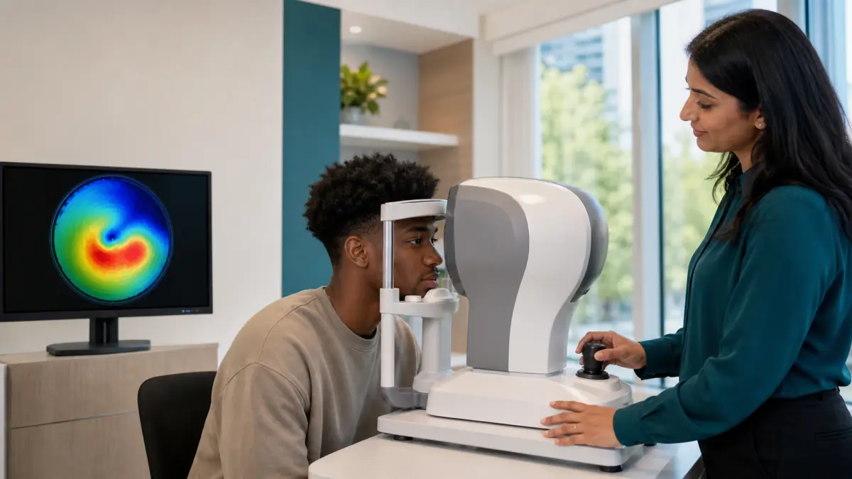
Corneal topography is a quick, non-contact test that maps the curvature of the front corneal surface. The patient rests the chin and forehead against the instrument, looks at a target, and keeps the eyes open for a brief capture. Nothing needs to touch the cornea, and the test does not use radiation.
The resulting colour map is not a photograph of damage. It is a coded representation of curvature. Depending on the instrument’s scale, warmer colours usually indicate steeper regions and cooler colours flatter regions. The clinician examines the pattern, symmetry, steepest area, astigmatism, and how the map relates to vision.
A striking colour does not diagnose keratoconus by itself. Map scales, tear-film quality, blinking, eyelid position, contact-lens wear, and capture alignment can affect results. Poor-quality scans should be repeated rather than overinterpreted.
Tomography and thickness measurements add depth
Topography primarily describes the front surface. Corneal tomography reconstructs the cornea in three dimensions and can assess the front and back elevation, curvature, and distribution of thickness. Pachymetry measures corneal thickness, while some tomography systems include thickness data in the same scan.
These measurements are useful because an early ectatic pattern may appear in the posterior surface or in the way the cornea thins before it is obvious during a slit-lamp examination. The thinnest point, its location, the rate at which thickness increases toward the periphery, and the relationship among several indices are considered together.
No single number settles every case. A naturally thin cornea is not necessarily keratoconus, and a measurement within an average range does not automatically exclude an early asymmetric condition. Diagnosis is a pattern-recognition process informed by the entire examination.
Serial maps reveal whether the cornea is changing
One scan is a snapshot. Two or more reliable scans taken over time can show whether steepness, elevation, thickness, prescription, or corrected vision is changing. This distinction is central because the decision to recommend corneal cross-linking often depends on evidence of progression, not simply the presence of an unusual map.
Comparison works best when scans are captured with the same instrument and under similar conditions. The clinician may ask a contact-lens wearer to leave lenses out before mapping because lenses can temporarily alter corneal shape. The required interval depends on lens type, fit, wear time, and the purpose of the test; follow the clinic’s specific instructions rather than choosing an arbitrary number of days.
Keep copies of previous prescriptions, maps, and specialist reports. If you move or change clinics, a historical scan may be more valuable than a single new result because it establishes the rate and direction of change.
What a keratoconus assessment may include
Corneal imaging is only one part of the visit. A thorough assessment may include:
- questions about the timing of vision changes, glare, ghosting, rubbing, allergy, and family history;
- uncorrected and corrected visual acuity in each eye;
- measurement of the current glasses prescription;
- retinoscopy to observe the pattern of light reflected from the eye;
- keratometry to estimate the main corneal curves;
- slit-lamp examination for thinning, stress lines, scarring, or other corneal findings;
- corneal topography or tomography;
- pachymetry or another thickness measurement;
- assessment of the tear film and eyelids; and
- a dilated retinal examination when indicated to exclude other causes of reduced vision.
Testing is individualized. A person with a stable early pattern may need periodic imaging, while a rapidly changing young patient may require closer follow-up and referral to a corneal ophthalmologist.
Clearer vision and disease stabilization are separate goals
Keratoconus care becomes easier to understand when divided into two questions:
- What will improve the quality of vision now?
- What may reduce the risk of the cornea becoming more irregular later?
Glasses and contact lenses address the first question. They change how light enters the eye but do not strengthen the cornea or reliably prevent progression. Corneal cross-linking addresses the second question. Its primary purpose is stabilization; it is not a guarantee that glasses or lenses will no longer be needed.
Some people need only updated correction and monitoring. Others need specialty contact lenses for useful vision, cross-linking for documented progression, or both. Treating one goal does not make the other irrelevant.
Glasses can work well in early disease
When irregularity is mild, glasses may provide comfortable, functional vision. A new prescription can correct the regular portion of nearsightedness and astigmatism even though it cannot neutralize every higher-order distortion.
Frequent prescription changes should prompt reassessment rather than an endless cycle of remaking lenses. If a technically accurate prescription still leaves shadows or glare, corneal mapping may explain why. Conversely, a stable prescription and stable maps may support continued observation.
Glasses remain useful even for specialty-lens wearers. They provide backup during infections, allergy flares, lens damage, or the hours before and after lens wear.
Contact-lens options are tailored to the cornea
Contact lenses can create a more regular optical surface in front of an irregular cornea. The appropriate design depends on disease severity, corneal shape, comfort, ocular-surface health, handling ability, visual needs, and cost.
Options may include:
- Soft or soft toric lenses: suitable for some mild cases, especially when most blur is still regular.
- Custom soft keratoconus lenses: thicker or specially designed optics may mask more irregularity than a standard soft lens.
- Rigid gas-permeable lenses: a small rigid surface can provide clear optics, but alignment, movement, edge position, and corneal clearance require careful fitting.
- Hybrid lenses: a rigid centre is surrounded by a soft skirt, combining rigid optics with a different comfort and handling profile.
- Piggyback systems: a rigid lens is worn over a soft carrier lens in selected cases.
- Scleral lenses: a larger rigid lens rests on the white sclera and vaults over the cornea, holding a fluid reservoir between lens and eye.
No design is universally best. A lens that produces excellent chart vision but causes excessive pressure, poor oxygen delivery, redness, or unstable wear is not a successful fit. Regular follow-up checks the cornea, lens surfaces, solution routine, and whether the fit remains appropriate as shape changes.
Specialty lenses improve vision only while they are worn. They do not permanently press the cornea back into shape and should not be treated as a substitute for progression monitoring. Do not sleep, shower, or swim in lenses unless your eye-care professional has given specific instructions; water exposure raises infection risk.
Corneal cross-linking aims to slow progression
Corneal collagen cross-linking uses riboflavin and controlled ultraviolet-A light to strengthen bonds within corneal tissue. It is considered when keratoconus is progressing and the eye meets safety and candidacy requirements. The aim is to slow or stop further weakening and shape change.
This treatment is not primarily a vision-correction procedure. Some people experience a change in corneal shape or prescription afterward, but sharper unaided vision is not guaranteed. Glasses or contact lenses are commonly still needed. The realistic success measure is often a stable cornea over time.
Cross-linking protocols differ. In the widely used epithelium-off approach, the thin surface layer of the cornea is removed before riboflavin and ultraviolet treatment. Epithelium-on or transepithelial approaches leave that layer in place, but protocol availability and evidence differ. A corneal specialist should explain the recommended technique, benefits, limitations, recovery, and alternatives for the individual eye.
Candidacy can depend on documented progression, age, corneal thickness, scarring, current vision, surface health, pregnancy or health considerations, and the treatment system being used. Possible risks include pain during early healing, light sensitivity, haze, infection, scarring, delayed surface healing, and loss of vision. These details require a surgical consultation rather than a generic online recommendation.
After cross-linking, follow-up imaging remains important. Stabilization is judged over time, and the prescription or contact-lens fit may need to be reassessed after the cornea heals.
Ring segments and corneal transplant are selected options
Intracorneal ring segments are small arcs placed within the cornea in selected mild-to-moderate cases. They can alter corneal shape, reduce some irregularity, and sometimes make contact-lens fitting easier. They do not cure the underlying tissue weakness and are not suitable for every cone location, thickness, or level of scarring. They may be discussed alongside cross-linking because reshaping and stabilization serve different purposes.
Advanced keratoconus with substantial scarring, extreme thinning, poor vision despite correction, or inability to tolerate lenses may require a corneal transplant. Depending on the eye, a surgeon may replace a deep portion of the cornea while retaining its innermost layer or perform a full-thickness transplant.
Transplantation can restore a clearer and more regular optical window, but it is not an instant return to uncorrected vision. Healing is lengthy, astigmatism can remain, and glasses or rigid lenses may still be needed. Infection, rejection, wound problems, and other surgical complications require long-term specialist care. Most people with keratoconus are managed without reaching transplantation, particularly when progression is recognized and treated appropriately.
Keratoconus changes refractive-surgery decisions
LASIK removes and reshapes corneal tissue. A cornea with keratoconus is already biomechanically weak, so routine LASIK is not appropriate and may worsen ectasia. This is one reason reputable refractive-surgery screening includes corneal topography or tomography rather than relying only on prescription and thickness.
Do not conceal a family history or old keratoconus concern during a laser-vision consultation. Even a subtle or “suspect” map can materially change the risk assessment. Some highly selected patients may hear about customized surface treatments combined with stabilization, but these are specialist decisions with specific limitations—not ordinary laser vision correction and not a universal way to eliminate lenses.
Sudden cloudy vision can indicate acute hydrops
Keratoconus usually changes gradually, but a rare complication called acute corneal hydrops can appear suddenly. A break in an internal corneal layer allows fluid to enter the cornea, producing abrupt clouding, marked blur, discomfort or pain, watering, and light sensitivity.
Sudden cloudy vision in an eye with known or suspected keratoconus needs same-day urgent eye care. Do not rub the eye, insert a contact lens, or drive yourself if vision is impaired. Severe pain, injury, chemical exposure, or sudden vision loss may require emergency-department care. Optima Eye Care offers emergency eye-care guidance for North York patients, but an online booking should never delay immediate care for a true emergency.
Practical steps while awaiting assessment
Avoid rubbing or pressing the eyes. If allergy or dryness drives the urge, ask which preservative-free lubricants, cool compresses, or allergy therapies are appropriate for you. Do not use another person’s drops or repeatedly use redness-relief drops to mask symptoms.
Bring your current glasses, contact lenses, lens packaging, solution names, and older prescriptions to the visit. Note approximately when night glare, ghosting, or one-sided blur began. Ask relatives whether anyone has had keratoconus, cross-linking, specialty hard lenses, or a corneal transplant.
If you wear contacts, ask the clinic in advance whether they must be removed before mapping and for how long. Continue any prescribed medication unless the clinician who prescribed it advises otherwise. Do not stop a medically necessary lens or medication based only on general web information.
Keratoconus assessments in North York
Changing astigmatism does not always mean keratoconus, and an unusual topography map does not tell the whole story. A useful evaluation combines the prescription, corrected vision, slit-lamp findings, corneal shape, thickness, family history, symptoms, and change over time.
When keratoconus is present, the plan should answer both central questions: how to provide the clearest practical vision today and how to protect the cornea from avoidable progression. Glasses or specialty lenses may address optical quality. Serial mapping establishes stability. A corneal referral can determine whether cross-linking or another procedure is appropriate.
If your prescription keeps changing, one eye produces ghost images, or keratoconus runs in your family, request an appointment with Optima Eye Care for a North York assessment. Bring previous prescriptions or scans if you have them. Earlier comparison gives the clinician a better chance to distinguish a naturally unusual cornea from one that is actively changing.