

Eye Care Resources
Corneal Abrasion in North York: What to Do After Scratching Your Eye
June 25, 2026
A fingernail catches the eye while you are picking up a child. A branch brushes your face during yard work. A contact lens feels wrong after a long day, or a grain of grit blows under the eyelid near a North York construction site. The eye begins watering, light feels painfully bright, and every blink feels as though something is still trapped beneath the lid.
These symptoms may come from a corneal abrasion—a scrape in the surface layer of the clear cornea at the front of the eye. An abrasion may be microscopic and still cause disproportionate pain because the cornea is densely supplied with nerves. It may also resemble a retained foreign body, infection, chemical injury, or deeper penetrating wound. Appearance in the mirror cannot reliably separate them.
Prompt assessment is important when pain, light sensitivity, redness, or blurred vision follows an eye injury. This is especially true for contact-lens wearers, injuries involving metal or plant matter, high-speed particles, and anyone whose vision is reduced. Optima Eye Care offers emergency eye-care guidance for North York patients, while suspected penetrating injuries, severe chemical exposure, or major trauma require emergency-department care.
The corneal epithelium is a protective surface
The cornea is the transparent front window that helps focus light into the eye. Its outer epithelium is a thin, tightly joined layer that acts as a barrier against germs, debris, and fluid. A corneal abrasion occurs when part of this surface layer is scraped or lifted away.
Because the cornea has many sensory nerve endings, an exposed area can produce sharp pain, burning, tearing, and reflex eyelid squeezing. A larger scratch is not always the most painful, and pain level alone does not show how deep an injury is. A small abrasion near the centre may blur vision more than a larger peripheral one because it lies directly across the visual axis.
Most uncomplicated surface abrasions heal quickly as epithelial cells slide and divide to cover the defect. Healing does not eliminate the need for an initial examination when the cause or depth is uncertain. Germs can enter through the disrupted barrier, retained material can remain beneath a lid, and a wound that looks superficial at home may be more serious.
Common causes range from ordinary to high risk
Corneal abrasions can follow:
- a baby’s finger, adult fingernail, or pet paw;
- a paper or cardboard edge;
- a twig, leaf, gardening debris, or sawdust;
- dust, sand, grit, or cosmetic particles;
- a makeup brush, mascara wand, or false-eyelash tool;
- inserting, removing, or overwearing a contact lens;
- rubbing an eye while something is trapped under the lid;
- a broken or chipped contact lens;
- sports contact or a toy;
- improper use of a towel or tissue near the eye; or
- metal, stone, glass, or wood fragments generated by tools.
The mechanism changes the level of concern. A clean paper edge moving slowly is different from metal striking metal, grinding, drilling, hammering, mowing, or using compressed air. High-speed particles can penetrate the eye even when the outside injury looks small. Plant matter and soil can introduce unusual organisms. Contact-lens wear increases concern for aggressive bacterial infection.
Tell the clinician exactly what happened, including the material, speed, protective equipment, time of injury, contact-lens use, and any attempt to remove the object. Those details shape the examination and treatment.
What does a scratched cornea feel like?
Typical symptoms include:
- sudden sharp, burning, or stinging pain;
- a gritty or foreign-body sensation;
- heavy tearing;
- redness;
- light sensitivity;
- blurred or hazy vision;
- difficulty keeping the eye open;
- frequent blinking or eyelid spasm;
- headache caused by pain and light sensitivity; or
- swollen eyelids after prolonged irritation.
Symptoms often worsen with blinking because the upper eyelid moves across the injured surface. A scratch under the upper lid or a particle trapped there can create a repeated vertical pattern on the cornea each time the person blinks.
Not every abrasion begins at the exact moment of trauma. A person may remove a lens at night and wake with pain hours later. Another may feel improved after rinsing but have symptoms return when the eye opens the next morning. A delayed or recurring pattern can provide an important diagnostic clue.
An abrasion and a foreign body can feel identical
The sensation that something remains in the eye may persist after a particle has washed away because the exposed corneal nerves continue sending a foreign-body signal. Alternatively, a speck may still be lodged under the eyelid or embedded in the cornea.
Do not keep rubbing, sweeping the eye with a cotton swab, or asking someone to probe beneath the lid. Those actions can enlarge a scratch or push material more deeply. An optometrist or physician can evert the eyelid safely, inspect the surface under magnification, and remove superficial material with appropriate instruments when indicated.
An embedded object, an object visibly protruding from the eye, or material that prevents the eyelids from closing must be left in place. Do not pull it out and do not press a patch against it. Protect the eye from further contact with a rigid shield if one is available and go to an emergency department.
For detailed guidance about loose and embedded material, see our foreign object in the eye article.
Safe first aid begins with clean hands and gentle irrigation
If the injury appears to involve loose dust or grit rather than penetration or chemical exposure:
- Stop the activity and move away from ongoing dust or debris.
- Wash and dry your hands.
- Remove a contact lens if it comes out easily. Do not continue manipulating the eye if the lens seems stuck.
- Rinse with sterile saline or clean, lukewarm water. A workplace eyewash station is appropriate when available.
- Blink several times to encourage natural tearing.
- Keep the eye relaxed and arrange prompt assessment if pain, light sensitivity, redness, or blur remains.
Use a gentle stream. High-pressure water can worsen an injury. Do not add salt, baking soda, essential oil, or any other household ingredient to make your own eye solution. Bottled drinking water is not sterile, but clean water is preferable to delaying necessary irrigation when debris or a chemical needs to be flushed.
If a chemical entered the eye, irrigation takes priority over almost everything else. Begin flushing immediately with copious lukewarm water and continue while emergency advice is obtained. Remove contact lenses if they do not wash out, but do not delay irrigation to search for them. Bring the product container or safety data information without exposing anyone else to the chemical. Chemical injuries require emergency assessment even if the eye feels better after rinsing.
What not to do after scratching an eye
Avoid several common responses:
- Do not rub or press the eye.
- Do not use tweezers, needles, tissues, cotton swabs, or fingernails to remove material.
- Do not put a contact lens back in.
- Do not use redness-relief drops to disguise the symptoms.
- Do not use leftover antibiotic or steroid drops.
- Do not borrow another person’s medication.
- Do not apply essential oils, honey, breast milk, saliva, or homemade mixtures.
- Do not drive if pain, tearing, light sensitivity, or blur affects vision.
Do not tape the eye shut or improvise a pressure patch. Patching is not required for most simple abrasions and can be inappropriate when infection risk is elevated. A clinician may occasionally select a patch or therapeutic bandage lens for a specific injury, but those decisions include follow-up and infection precautions.
Contact-lens wear changes the urgency
A painful red eye in a contact-lens wearer must be treated cautiously. Contact lenses can produce a mechanical scratch, trap debris, reduce oxygen at the surface, or allow bacteria to contact a disrupted epithelium. Certain infections associated with lens wear can progress quickly and threaten vision.
Remove the lens and seek same-day assessment when possible, particularly for pain, light sensitivity, discharge, reduced vision, or a visible white spot. Do not restart lens wear simply because symptoms improve overnight. The clinician needs to confirm that the surface is closed, infection is absent, and the lens or care routine is not contributing.
Bring the lenses, case, packaging, and solution details if doing so does not delay care. Do not clean or discard everything before the visit when a clinician may want to know what was used. Never wear the affected lens again unless specifically told it is safe; disposable lenses and cases are often replaced after an injury or infection concern.
Sleeping, showering, swimming, or using tap water with contact lenses increases risk. “Topping off” yesterday’s solution does not disinfect a lens. Following replacement schedules and receiving regular contact-lens checks reduces avoidable complications but cannot prevent every accidental scratch.
Power tools and high-speed particles need emergency evaluation
Hammering metal, grinding, drilling, cutting tile, using a wire wheel, or working beneath a vehicle can launch particles faster than a blink reflex can protect the eye. A tiny metal fragment may lodge in the cornea and leave a rust ring, or it may penetrate more deeply.
Seek emergency assessment after a high-velocity mechanism even if discomfort seems mild. Do not rely on the fact that safety glasses were worn; open sides, gaps, and improper fit can still permit entry. Do not use a magnet near the eye or try to lift out metal yourself.
Warning signs of possible penetration include a misshapen pupil, marked vision reduction, blood inside the eye, fluid leaking from a wound, a visibly deep object, or an eye that looks structurally different. These signs may be absent. The mechanism alone can justify urgent imaging and ophthalmic evaluation.
Keep both eyes still as much as practical because they move together. Do not eat or drink while awaiting emergency advice in case a procedure requiring anesthesia is needed. Avoid all pressure on the injured eye.
Plant matter and fingernails have special implications
A leaf, twig, thorn, soil particle, or dirty fingernail can introduce organisms into a corneal defect. These injuries do not guarantee infection, but the material matters when a clinician selects medication and follow-up. Report gardening, compost, farm, or plant exposure clearly.
Fingernail abrasions can also disrupt how newly healed epithelial cells attach to the layer underneath. Weeks, months, or occasionally years later, a small area may loosen again when the eyelid opens after sleep. This is called recurrent corneal erosion.
Do not assume that pain returning after apparent healing means a new particle entered the eye. Recurrent erosion and infection can feel similar and both need assessment. A history of the original scratch helps the clinician interpret the pattern.
Fluorescein makes a surface defect visible
During the examination, visual acuity is measured first when it can be done safely. The clinician evaluates pupil shape and response, eye movement, eyelids, conjunctiva, cornea, and the depth of the front chamber of the eye.
Fluorescein is an orange-yellow diagnostic dye that glows bright green under cobalt-blue light wherever the surface epithelium is missing. It outlines the location, size, and pattern of an abrasion. The dye does not permanently stain a healthy cornea, although it can temporarily discolour tears, skin, or a soft contact lens.
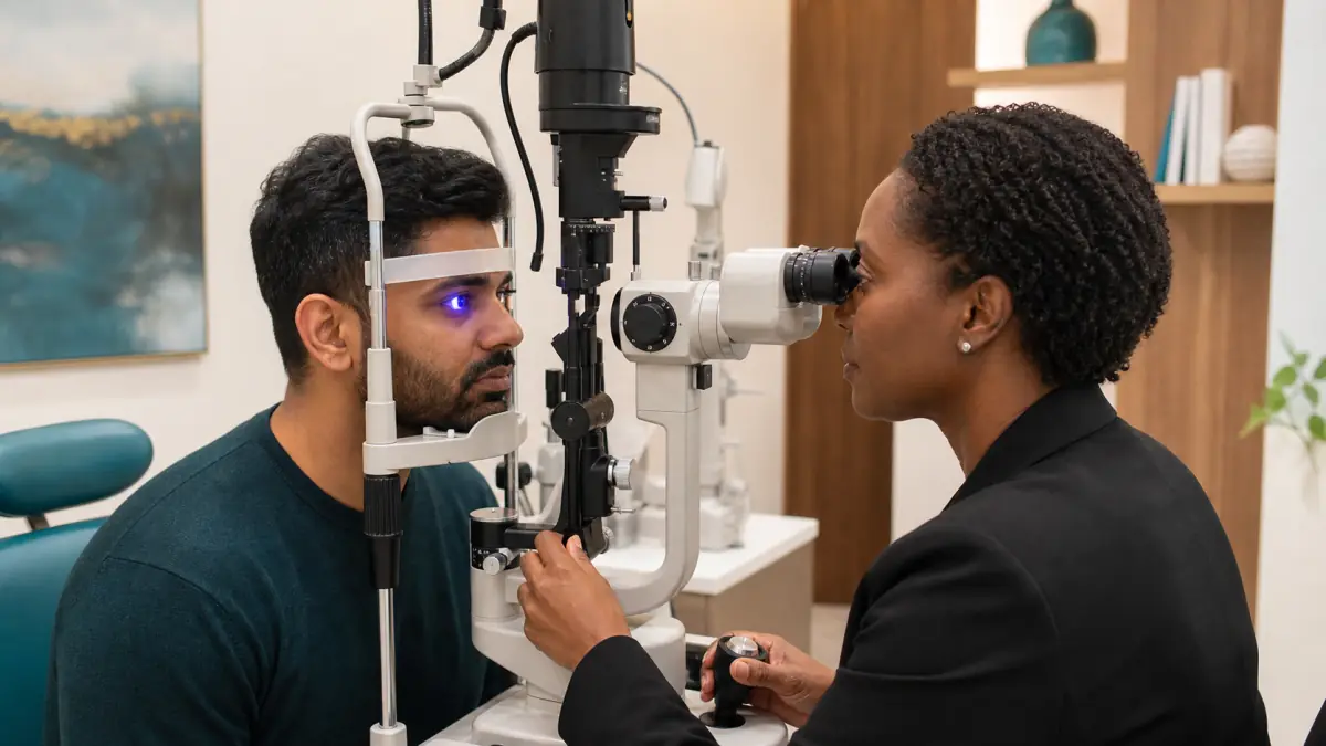
A slit-lamp microscope provides a magnified, illuminated view. The clinician may gently turn the upper eyelid to search for trapped material. A particular fluorescein flow pattern can raise concern that fluid is leaking through a full-thickness wound. If penetration is suspected, further manipulation, pressure measurement, and routine drops may be avoided while emergency ophthalmic care is arranged.
The examination also checks for a corneal infiltrate or ulcer, inflammation inside the eye, lens complications, and other causes of pain. A scratch cannot always be confirmed from a photograph because the epithelial defect may be transparent in ordinary light.
Treatment is matched to cause, size, and infection risk
After loose material is removed and the depth is established, care aims to protect healing tissue, reduce pain, and prevent infection. The exact regimen depends on whether the abrasion is simple, related to a contact lens, contaminated by plant or soil material, recurrent, or accompanied by another injury.
An antibiotic ointment or drop may be prescribed to reduce bacterial infection risk. Contact-lens wearers require different antimicrobial considerations than a person with a clean, non-lens-related scratch. Use the medication at the stated frequency and for the stated duration. Do not stop as soon as pain improves unless instructed.
Preservative-free lubricants or ointment may reduce friction. Oral pain relief may be recommended based on age, health conditions, allergies, pregnancy, kidney function, stomach history, and other medicines. Drops that temporarily relax the focusing muscle and dilate the pupil are sometimes used when internal spasm contributes to pain and light sensitivity.
A clinician may place a bandage contact lens for a selected abrasion. This is a medical device requiring antimicrobial coverage and close review, not an ordinary lens used as a bandage at home. It can mask worsening symptoms or promote infection if used inappropriately.
Numbing drops belong in the examination setting
Topical anesthetic drops can provide dramatic relief and allow a thorough examination, but repeated unsupervised use can delay healing, mask infection, and cause severe corneal damage. They should not be taken home or borrowed for recurring pain unless an ophthalmic specialist has provided a tightly controlled plan for a specific situation.
Pain returning after the diagnostic drop wears off is expected and does not mean the examination caused additional injury. Follow the recommended comfort plan and contact the clinic if pain is severe, increasing, or different from what was discussed.
Likewise, steroid drops should never be started after a scratch without examination. Steroids can worsen certain bacterial, fungal, or viral corneal infections and alter healing. A bottle prescribed for allergy or a previous inflammation is not safe simply because it is labelled for the eye.
Most simple abrasions improve quickly—but follow-up matters
Small uncomplicated abrasions often close within one or two days. Larger defects can take several days or longer. Comfort generally improves as the surface seals, but complete symptom relief and stable vision may lag behind initial closure.
Healing estimates are not deadlines. A contact-lens-related defect, contaminated injury, diabetes, severe dry eye, abnormal eyelid closure, reduced corneal sensation, or underlying corneal disease can slow recovery. A central abrasion may continue to affect vision even while the edges are healing.
Attend the recommended review even if the eye feels better. Follow-up can confirm that the epithelium has closed, no infiltrate has developed, and the medication can be stopped or changed safely. Seek earlier reassessment if pain, redness, discharge, light sensitivity, or blur increases.
A corneal ulcer can begin like a scratch
Microbial keratitis is an infection of the cornea. It may develop after trauma, contact-lens wear, or contamination and can produce a focal white or grey spot, increasing pain, discharge, marked light sensitivity, and reduced vision. It can progress beyond the surface and leave permanent scarring.
Do not wait for a scheduled visit if symptoms are worsening rather than improving. An abrasion is expected to trend toward comfort as it heals; escalating pain or a new visible spot deserves urgent evaluation.
Herpes simplex can also create a branching epithelial defect that may initially be described as a “scratch,” even without trauma. Treatment differs and steroid use can be dangerous in certain active infections. Recurrent one-sided redness, pain, light sensitivity, cold-sore history, or unexplained previous episodes should be reported.
Recurrent corneal erosion causes pain on waking
In recurrent corneal erosion, a patch of epithelium does not adhere securely to its underlying layer. The closed eyelid can stick to that area during sleep; when the eye opens, the surface lifts again. Pain is often sudden on waking or during the night, accompanied by tearing and light sensitivity.
Previous fingernail, paper, or plant injuries can lead to this problem, but it may also occur with an inherited or age-related epithelial basement-membrane condition. Dry eye, eyelid inflammation, and incomplete nighttime closure may contribute.
Initial management may include intensive lubrication, especially at bedtime, or other clinician-directed therapy. Persistent episodes can require a bandage lens, hypertonic ointment, or a corneal procedure designed to improve epithelial attachment. Options have different risks and evidence, so treatment is based on frequency, location, severity, and examination findings.
Repeatedly treating morning pain with leftover antibiotics or numbing drops does not address the attachment problem. Keep a record of timing, prior injury, and which eye is affected, then arrange assessment.
Children may not explain the symptoms clearly
A child with a corneal abrasion may keep one eye closed, resist bright light, rub the face, cry, or refuse to open the eye for examination. A parent may know that a toy, sibling’s finger, pet, or plant was involved but not whether an object remains.
Do not force the lids apart, hold a child down to probe the eye, or apply someone else’s drops. Gentle irrigation may be attempted for loose dust if the child can cooperate and there is no concern for penetration or chemical injury. Otherwise seek prompt medical care.
Any high-speed mechanism, sharp object, chemical, visibly distorted eye, severe swelling, or significant vision concern requires emergency evaluation. A calm history of what happened is often more useful than repeated attempts to inspect the eye at home.
Preventing the next injury
Wear safety eyewear appropriate to the task, with side protection when particles can travel around a simple front lens. Regular glasses are not a substitute for certified protective eyewear. Grinding, hammering, chemical work, and high-impact sport may require goggles or a face shield in addition to glasses.
Trim children’s fingernails, supervise sharp toys, and keep branches and leaves away from face level during yard work. Pause before opening a package, applying mascara, or handling a lens when someone may bump your arm.
Contact-lens wearers should wash and dry hands, keep nails away from the cornea, replace damaged lenses, and never force a lens that feels folded or stuck. Seek fitting advice when removal repeatedly scratches the eye or a lens rides under the upper lid.
Workplace injuries should be reported according to the employer’s safety process, but paperwork must not delay first aid or emergency assessment. Review how the injury bypassed the protective equipment so the same pathway can be corrected.
Corneal abrasion care in North York
A scratched cornea can be intensely painful, yet the same symptoms can also represent a trapped particle, corneal infection, chemical burn, or penetrating injury. A slit-lamp examination with fluorescein identifies the surface defect, checks beneath the eyelids, and looks for complications that cannot be judged at home.
Optima Eye Care can assess suspected surface injuries for North York patients and coordinate referral when a wound is deep, infected, contaminated, or otherwise beyond routine management. If the eye was exposed to a chemical, struck by a high-speed particle, penetrated by an object, or has severe pain or vision loss, go directly to emergency care.
For persistent pain, tearing, light sensitivity, or blur after a minor scratch, request urgent guidance from Optima Eye Care and explain the injury mechanism when you call. Remove contact lenses, do not rub the eye, and do not use leftover drops while waiting for professional advice.